
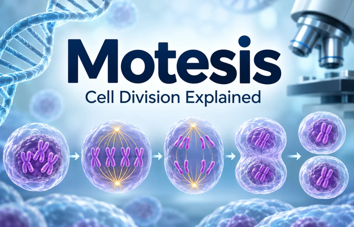
Motesis is not a recognized standard term for a biological process. In the context of cells and human biology, the closest established term is mitosis: the carefully controlled process in which a cell separates its duplicated chromosomes into two nuclei, usually followed by division into two daughter cells. Mitosis enables growth, tissue maintenance, and the replacement of many worn-out cells.
If you searched for “Motesis” because you heard the word in a science lesson or saw it written online, mitosis may be what you need. I’ll explain the distinction, show what happens inside a dividing cell, and separate established facts from common misconceptions. The spelling matters because a single changed letter can lead you away from trustworthy medical and biology references.
What Does Motesis Mean?
As of November 2026, I could not verify “Motesis” as a defined biological term in major authoritative references. The National Institute of General Medical Sciences and the National Human Genome Research Institute both document mitosis, not “Motesis,” when describing ordinary cell division.
That does not rule out Motesis as a brand or coined name. It simply means I cannot assign it a universal definition.
Motesis may be the spelling you encountered; mitosis is the established scientific concept. Check the original context before equating them.
Is Motesis simply a misspelling of mitosis?
It could be, especially when the surrounding text mentions DNA, chromosomes, cell growth, or biology classes. However, I would not assume that every occurrence is a typo.
Context is the deciding factor. A biology textbook pointing to “Motesis stages” is probably referring to mitosis; a company logo using the same letters could mean something completely different.
Why Mitosis Is Essential for Life
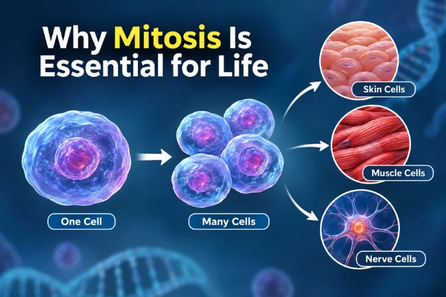
Mitosis is sometimes called the body’s copying system, but the challenge is distribution. Each new cell needs the genetic instructions required to function.
Growth from one cell to many
Human development begins with a fertilized egg. Repeated rounds of cell division help form the enormous number of cells that make up the body.
As development continues, cells also specialize. A skin cell and a muscle cell may contain essentially the same DNA, yet use different genes to perform different jobs.
Mitosis does not, by itself, determine what a new cell will become. It supplies new cells; developmental signals and gene regulation guide their roles.
Replacing cells and repairing tissues
I find it helpful to picture the lining of the digestive tract: its cells experience constant wear and must be replenished. Skin and other tissues also depend on controlled cell production.
Not every tissue renews at the same rate, though. Some mature nerve cells rarely divide, if at all.
When a minor injury heals, cell division can help replace lost tissue, alongside inflammation, cell movement, and rebuilding of the surrounding structure. Saying that mitosis alone “heals wounds” would oversimplify a coordinated biological process.
The Cell Cycle: What Happens Before Division?
Here is a detail that frequently gets overlooked in explanations of Motesis or mitosis: DNA copying happens before mitosis, during a stage called S phase. Mitosis is mainly about safely separating chromosomes that have already been duplicated.
The complete sequence is called the cell cycle. It includes interphase, when the cell grows and prepares, and the M phase, when chromosomes separate and the cell typically divides.
The NHGRI cell-cycle glossary describes this recurring pattern.
| Cell-cycle stage | Main activity | Why it matters |
|---|---|---|
| G1 phase | The cell grows and carries out normal work | Builds resources for a possible division |
| S phase | DNA is replicated | Produces a copy of each chromosome |
| G2 phase | The cell prepares for division and checks readiness | Helps reduce errors before mitosis |
| M phase | Chromosomes separate; cytokinesis usually follows | Produces two daughter cells |
| G0 state | Some cells pause or leave the active cycle | Explains why all cells do not divide continuously |
Think of the process as preparing two complete sets of instructions before distributing them. If the instructions are not copied correctly, a tidy-looking division cannot guarantee a healthy result.
Another key point: not all cells proceed through this cycle at the same speed. The timing depends on cell type, tissue needs, growth signals, and conditions. There is no single universal countdown for all human cells.
The Stages of Mitosis Explained
Many classrooms summarize mitosis with four main stages: prophase, metaphase, anaphase, and telophase.
More detailed explanations also separate prometaphase and describe cytokinesis afterward. Both approaches can be useful as long as the underlying events are clear. The NIGMS glossary outlines the more detailed sequence.
1. Prophase: Chromosomes become visible
Before division, genetic material is relatively spread out in the nucleus. As prophase begins, it condenses into recognizable chromosomes.
Each copied chromosome consists of two sister chromatids joined at a specialized region called the centromere.
At the same time, the cell builds a mitotic spindle, a framework made largely of microtubules. The spindle will help move chromosomes to the correct destinations.
2. Prometaphase: The spindle makes contact
The nuclear envelope breaks down in typical animal-cell mitosis, allowing spindle microtubules to interact with the chromosomes.
Protein structures called kinetochores form at centromeres and provide attachment sites.
I picture this stage as securing packages before shipping: each chromosome must attach so its sister chromatids can travel toward opposite sides.
3. Metaphase: Chromosomes line up
The copied chromosomes align near the cell’s center, often called the metaphase plate. Their positions reflect tension from spindle fibers attached on opposite sides.
Metaphase is recognizable in microscope images, making it useful for studying chromosomes.
4. Anaphase: Sister chromatids separate
Once the cell is ready, the connections holding sister chromatids together are released. The separated chromatids move toward opposite poles. Each is now considered an individual chromosome.
This is the central distribution event. Ideally, each future nucleus receives one copy of every chromosome.
A mistake here may leave one daughter cell with too many chromosomes and the other with too few.
5. Telophase: Two nuclei take shape
At opposite sides of the cell, the separated chromosome groups begin to loosen. New nuclear envelopes form around them, and the spindle is dismantled as the cell completes nuclear division.
Two nuclei have formed, but two separate cells may not exist yet.
6. Cytokinesis: The cell physically divides
Cytokinesis separates the cytoplasm and partitions the cell into two. Animal cells generally constrict through a contractile ring, while plant cells build a cell plate that develops into a new dividing wall.
Strictly speaking, mitosis is nuclear division and cytokinesis is cytoplasmic division.
In everyday teaching, both are often grouped together as the final events of cell division. Knowing the distinction prevents confusion later.
What Actually Happens to Chromosome Numbers?
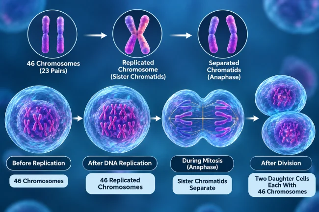
For human body cells, the familiar number is 46 chromosomes, arranged in 23 pairs. Before DNA replication, a typical diploid cell has 46 chromosomes.
After S phase, it still has 46 replicated chromosomes, but each consists of two sister chromatids.
That is the tricky part. Copying DNA does not automatically mean we should call the cell’s chromosome count 92. Chromosomes are usually counted by centromeres.
Once sister chromatids split in anaphase, each is recognized as a chromosome, and the dividing cell briefly contains two sets heading toward opposite ends.
After successful division, each daughter cell normally receives 46 chromosomes. I use this distinction whenever a simple diagram makes it look as though chromosomes appear or disappear by magic.
Chromosome numbers differ across species; mitosis does not give every organism 46.
Motesis, Mitosis, and Meiosis: What Is the Difference?
A search for Motesis can also lead to another similar-sounding biology term: meiosis. These are not interchangeable processes.
Mitosis generally supports the production of new body cells. Meiosis makes cells with a reduced chromosome set for sexual reproduction.
The NHGRI meiosis definition and NIGMS comparison explain the difference.
| Feature | Mitosis | Meiosis |
|---|---|---|
| Main purpose | Growth and maintenance; some forms of asexual reproduction | Formation of haploid reproductive cells |
| Divisions after DNA replication | One | Two |
| Typical resulting cells | Two | Four |
| Human chromosome count per resulting cell | Normally 46 | Normally 23 |
| Genetic similarity | Usually very similar to parent cell | Genetically varied |
| Key mechanism | Sister chromatids separate | Homologous chromosomes separate first; sister chromatids separate second |
One subtlety deserves attention: mitosis does not make daughters absolutely identical under every circumstance.
DNA copying errors, chromosome segregation mistakes, and other changes can introduce differences. “Genetically identical” describes the normal outcome, not an unconditional guarantee.
How Cells Keep Division Under Control
Cells have ways to decide whether division should proceed. These controls matter because accurate chromosome separation begins long before metaphase.
Checkpoints and repair mechanisms
Cell-cycle checkpoints monitor conditions such as DNA integrity, replication progress, and spindle attachment.
When problems are detected, the cell may pause, attempt repair, or stop progressing. Proteins involved in these decisions are part of a tightly coordinated regulatory network.
For example, the spindle assembly checkpoint helps prevent anaphase from starting before chromosomes are properly attached.
It is not a conscious safety inspection. It is a set of molecular signals that respond to attachment and tension.
Why the safeguards are not perfect
Even with these controls, mistakes can occur. A chromosome may fail to separate properly, or a copied DNA sequence may acquire a change.
Whether that change matters depends on what happened, which cells are affected, and how the body responds.
This is a more accurate picture than portraying healthy cells as error-free machines or treating every abnormal division as cancer. Biology includes safeguards, exceptions, and varying consequences.
What Can Go Wrong During Mitosis?
Problems in chromosome distribution may create cells with an unusual number of chromosomes, a condition called aneuploidy.
Some affected cells stop dividing or die. Others persist, sometimes with consequences for tissue function.
An error that happens after fertilization can also produce mosaicism: different groups of cells within one person may carry distinct genetic information.
The NHGRI description of mosaicism makes clear that this does not always cause disease.
The connection between cell division and cancer
Cancer develops when normal controls over cell growth and survival are disrupted.
Changes in genes involved in growth signaling, DNA repair, and cell-cycle regulation can contribute. Some cancer cells continue dividing despite signals that would stop a healthy cell.
Still, mitosis itself is not a disease. It is necessary for ordinary life. The problem is uncontrolled or abnormal regulation, often alongside other genetic changes.
The National Cancer Institute explains how cancer cells differ from normal cells.
I would also avoid claiming that one random mitotic error automatically causes cancer. Cancer usually develops through a more complex accumulation of changes, and many abnormal cells never form tumors.
Five Misunderstandings Worth Clearing Up
“Motesis” is a separate medical procedure
There is no verified standard biology procedure under that name. If you encountered the spelling in a health-related post, check whether the author intended to say mitosis before relying on the explanation.
Every cell divides all the time
Cell division rates vary widely. Some cells renew frequently, while others remain inactive or highly specialized for long periods. Cell-cycle regulation is precisely what prevents constant, unnecessary multiplication.
Mitosis creates genetic diversity like meiosis
Mitosis ordinarily preserves genetic information. Meiosis includes mechanisms such as recombination and independent assortment that generate varied reproductive cells. Both involve chromosomes, but their outcomes are different.
DNA is copied during metaphase
DNA replication occurs in S phase, ahead of mitosis. Metaphase is when previously duplicated chromosomes align. Mixing up those events makes the whole sequence harder to follow.
More cell division always means better healing
Extra cell growth is not automatically beneficial. Healing requires signals that tell cells when to divide, migrate, specialize, and stop. Uncontrolled growth can damage normal tissue rather than restore it.
A Practical Way to Recognize Mitosis in a Diagram
When I inspect a cell-division illustration, I ask three questions: Are the chromosomes lined up, moving apart, or enclosed inside new nuclei?
Those clues usually identify the stage faster than memorizing a long list of labels.
If X-shaped replicated chromosomes are aligned across the center, the illustration is likely showing metaphase. If the sister chromatids have separated and are traveling to opposite ends, it is anaphase.
If two nuclei are re-forming, look for telophase. A visible pinch or forming cell plate points toward cytokinesis.
Then I check what happened beforehand. Were chromosomes copied before the diagram begins? Does the example show one division or two?
Those details help distinguish mitosis from meiosis even when the drawings look similar.
For Motesis claims online, examine the context and look for an established reference rather than trusting repetition.
The Takeaway
Motesis has no verified standard biological definition in the sources I reviewed, but mitosis is a well-established and essential form of nuclear division.
It works within the wider cell cycle, preserves chromosome information in normal circumstances, and supports growth and tissue renewal. Understanding where DNA replication, chromosome separation, and cytokinesis fit is the easiest way to make sense of the subject.
If you are studying this topic, begin with the official NIH explanations linked above and compare them with your textbook diagrams. If you encountered Motesis as a product or name, seek its original source rather than assuming it refers to biology.
Frequently Asked Questions About Motesis
What is Motesis in biology?
Motesis is not an established biology term. If the context involves dividing cells, the intended word may be mitosis.
What is the main purpose of mitosis?
Mitosis distributes duplicated chromosomes into two nuclei, supporting growth, maintenance, and the formation of new body cells.
What are the main stages of mitosis?
The traditional four are prophase, metaphase, anaphase, and telophase; detailed accounts also identify prometaphase and subsequent cytokinesis.
Is mitosis the same as meiosis?
No. Mitosis generally produces two daughter cells with the same chromosome number; meiosis produces haploid reproductive cells.
Can mistakes during mitosis cause disease?
Some errors can affect cell function and contribute to disease, but many are corrected or eliminated and not every error leads to cancer.
Disclaimer
This article is for educational purposes only. “Motesis” is not an established biological term and may refer to mitosis. The information is based on general scientific knowledge and should not replace professional medical advice or authoritative scientific references.

Claire Morgan is a health and wellness writer with 7 years of experience creating clear, research-informed content on nutrition, sleep, movement, preventive habits, and everyday wellbeing. She focuses on making reliable health information practical and easy to understand. Her articles are educational and do not replace professional medical advice.